toe joints anatomy
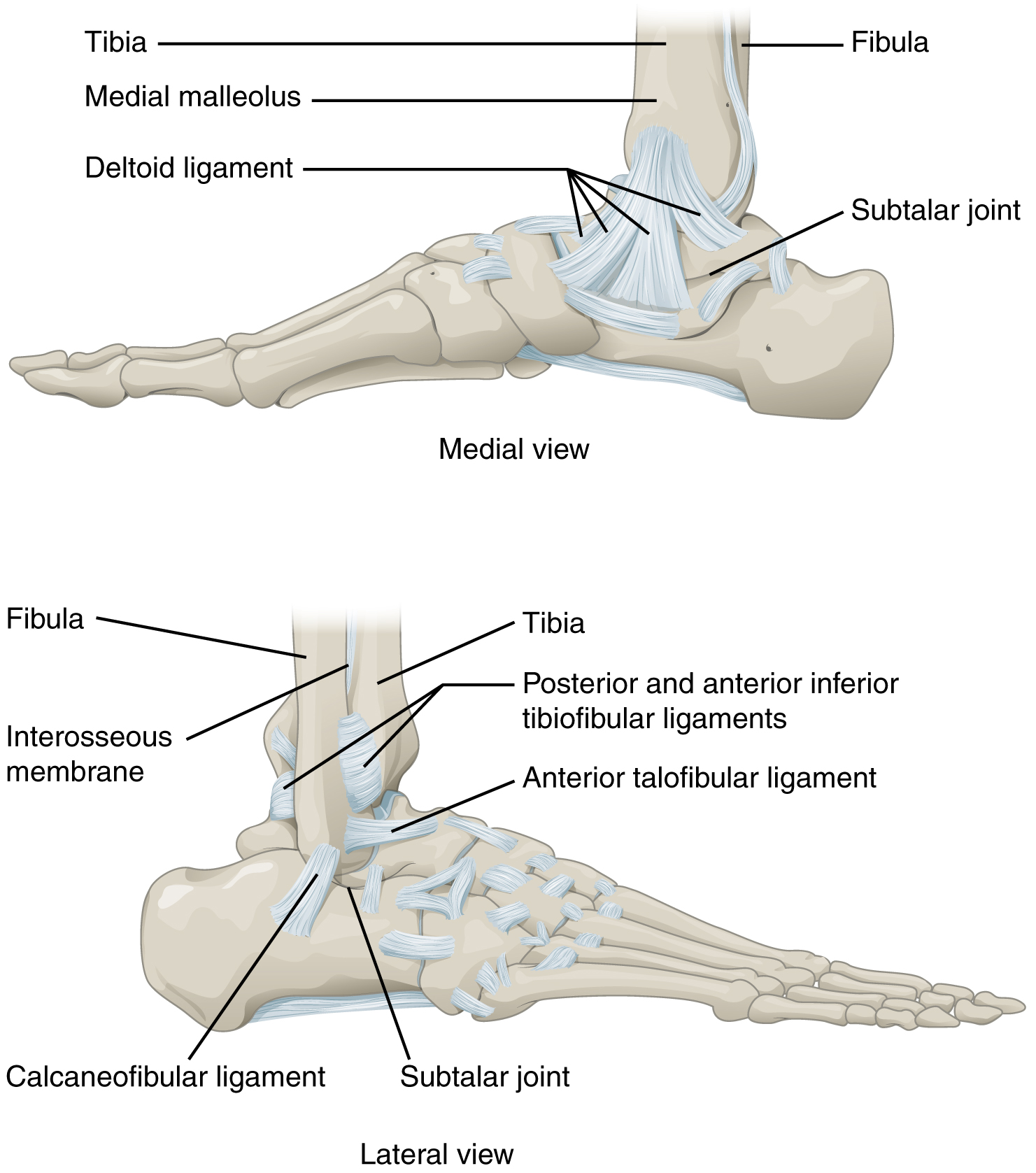
This figure shows the structure of the ankle and feet joints. The top. 13 Pictures about This figure shows the structure of the ankle and feet joints. The top : Foot Joints Photograph by Asklepios Medical Atlas, Ankle Foot Anatomy and also This figure shows the structure of the ankle and feet joints. The top.
This Figure Shows The Structure Of The Ankle And Feet Joints. The Top
 oerpub.github.io
ankle joint joints foot talocrural lateral bones talus medial eversion structure
Ankle Foot Anatomy
 anatomy.lexmedicus.com.au
foot bones anatomy toe joints ankle toes bone hallux phalanges inter metatarsals phalangeal through each another curling exception which
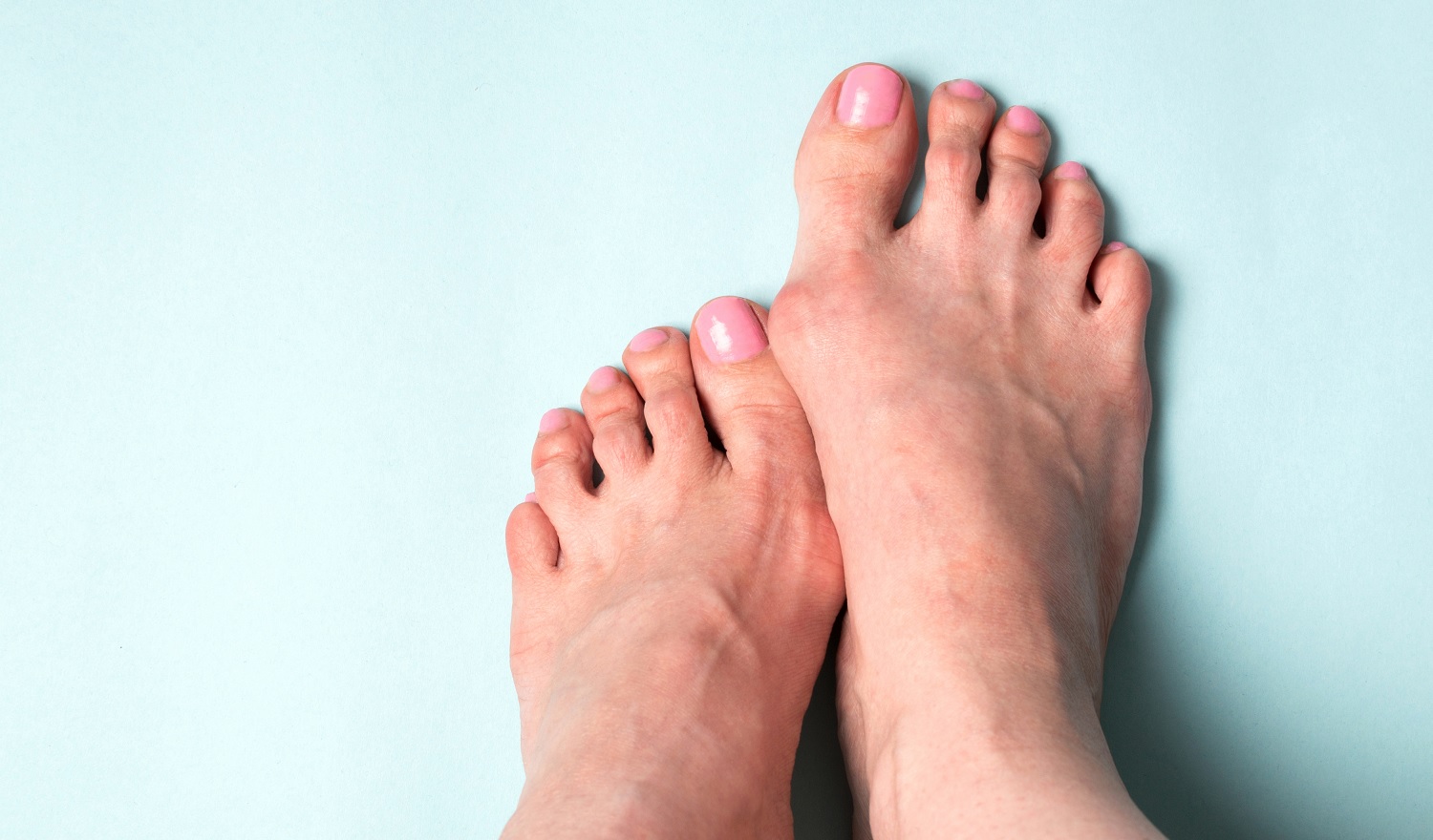
Hammer Time! The Difference Between Hammer Toes And Claw Toes
 footpoint.com.au
hammer toes claw difference between foot
Untitled Document [bio.sunyorange.edu]
![Untitled Document [bio.sunyorange.edu]](http://bio.sunyorange.edu/updated2/comparative_anatomy/anat_3/tibia,fibula_files/leg_all.jpg) bio.sunyorange.edu
bio.sunyorange.edu
foot leg alligator anatomy platypus tibia mink gator anat comparative fibula human updated2 sunyorange bio edu
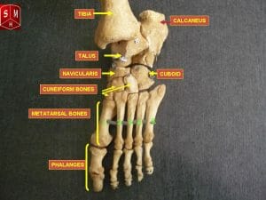
Foot (Anatomy): Bones, Ligaments, Muscles, Tendons, Arches And Skin
 biologydictionary.net
bones foot phalanges tarsus metatarsus bone file anatomy muscles navicular tendons ligaments skin
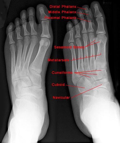
Foot X-ray - Normal Findings | Bone And Spine
 boneandspine.com
xray regions boneandspine
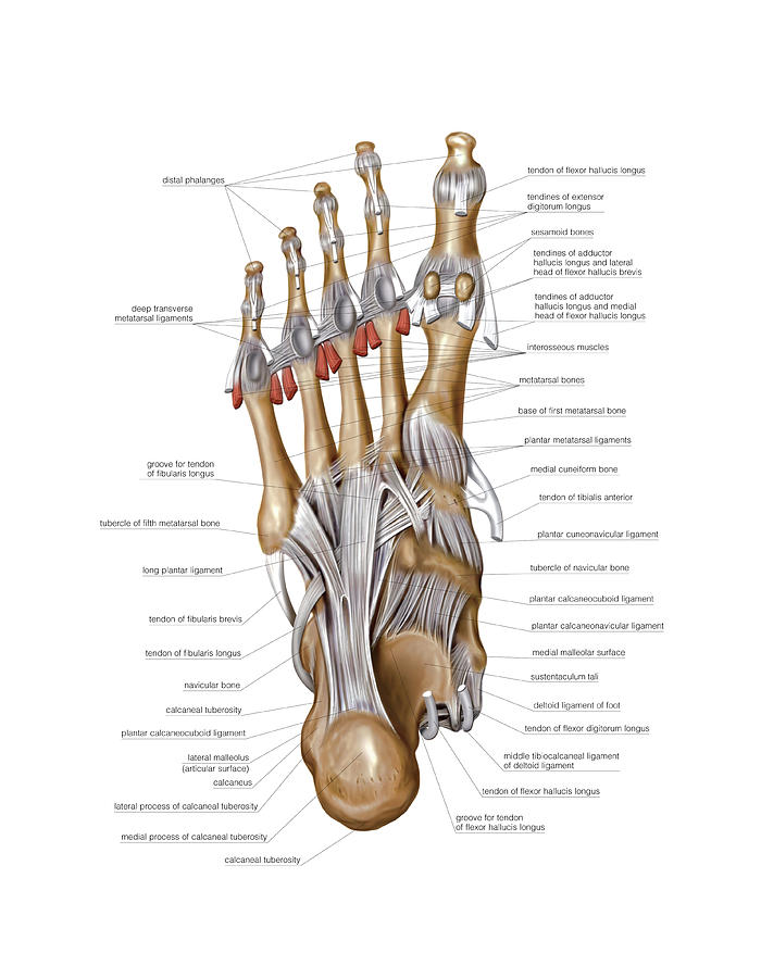
Foot Joints Photograph By Asklepios Medical Atlas
 fineartamerica.com
foot joints asklepios atlas medical anatomy photograph 1st uploaded august which
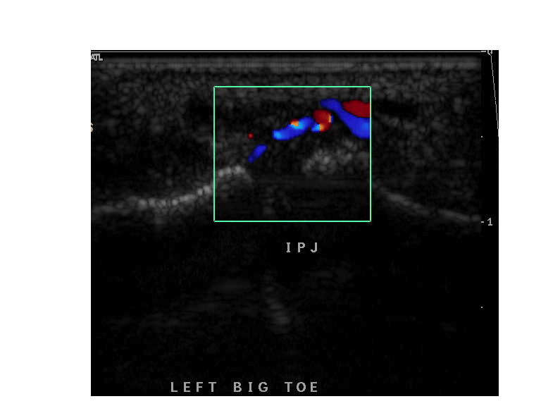
A Sonographer's Guide To Forefoot Imaging - Dr Iain Duncan
 driainduncan.com.au
toe joint forefoot synovitis ultrasound imaging guide fig ip sonographer
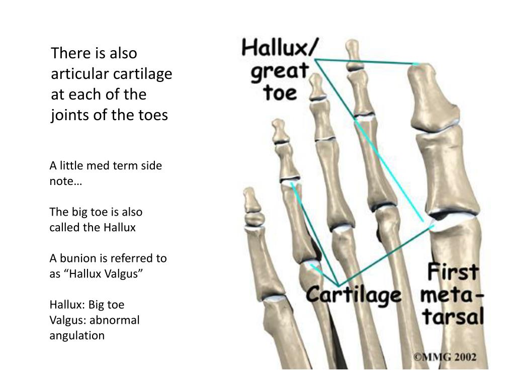
PPT - BONES OF THE FOOT & ANKLE PowerPoint Presentation, Free Download
 www.slideserve.com
foot ankle bones presentation
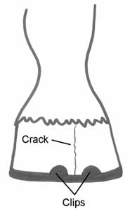
Joint, Tendon, Ligament, Bone, And Foot Problems
 www.infovets.com
e406 bone figure crack foot problems infovets
Gout | Image | Radiopaedia.org
 radiopaedia.org
gout radiopaedia tophi periarticular soft tissue arthropathy radiology podagra axial metatarsal case intraosseous crystals swelling erosions version
Foot Radiograph (an Approach) | Radiology Reference Article
 radiopaedia.org
foot radiograph radiopaedia approach normal radiology phalanges oblique figure
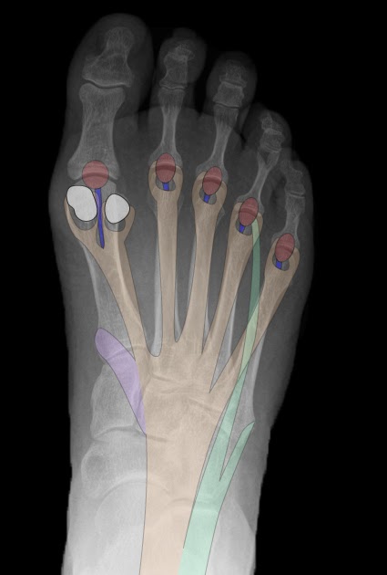
Roentgen Ray Reader: Plantar Aponeurosis: Anatomy
 roentgenrayreader.blogspot.com
plantar
Ankle foot anatomy. E406 bone figure crack foot problems infovets. Toe joint forefoot synovitis ultrasound imaging guide fig ip sonographer