sternal lymph node
Modern imaging lymph node staging of the head and neck region. 13 Pictures about Modern imaging lymph node staging of the head and neck region : Esophagectomy with Three-Field Lymph Node Dissection | CTSNet, Lymphoma in Dogs and also Extended Esophagectomy With 3-Field Lymph Node Dissection for.
Modern Imaging Lymph Node Staging Of The Head And Neck Region
 www.ejradiology.com
lymph p360 issue
An Unusual Cause Of Lymph Nodes Enlargement - The American Journal Of
 www.amjmed.com
lymph nodes calcified thoracic figure mediastinal enlargement unusual cause arrows transverse computed tomography scan mass section compressive bulky due amjmed
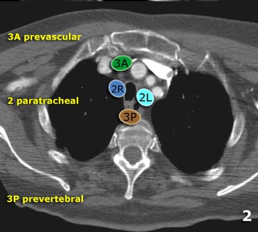
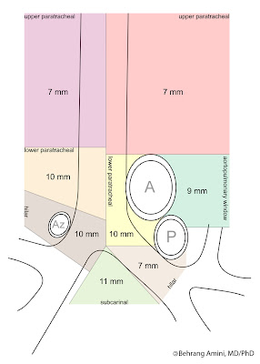
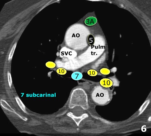
The Radiology Assistant : Mediastinum - Lymph Node Map
 radiologyassistant.nl
lymph nodes radiology mediastinum assistant mediastinal ct axial
Thoracic Wall - Atlas Of Anatomy
 doctorlib.info
anatomy axillary lymph nodes doctorlib info thoracic levels table lateral medical
Enlarged Limph Nodes In Hart Artery. Enlarged Lymph Nodes In Spleen
 pomo.se
neck nodes anatomy face cervical mri head node lymphatic lymph jugulodigastric enlarged occipital facial swollen imaging imaios area
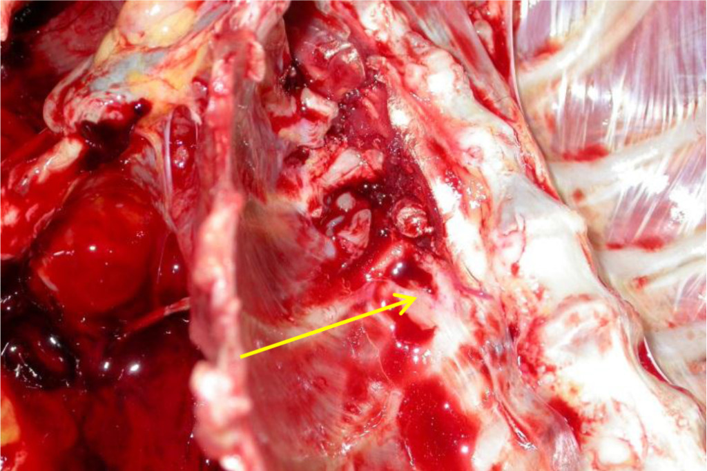
Researchopenworld.com
 researchopenworld.com
researchopenworld thoracic lymph intra aorta infiltrations vertebral
Lymphoma In Dogs
 www.slideshare.net
lymphoma lymphadenopathy splenomegaly
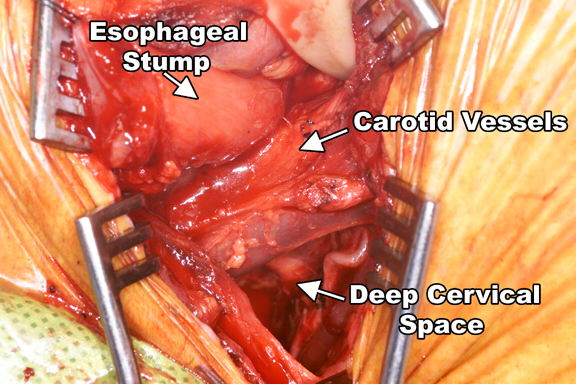
Esophagectomy With Three-Field Lymph Node Dissection | CTSNet
 www.ctsnet.org
lymph node dissection cervical esophagectomy carotid field three sheath posterior division ctsnet muscle sternal sternocleidomastoid adequate lateral exposure carried deep
Roentgen Ray Reader: Normal Limits For Thoracic Lymph Nodes
 roentgenrayreader.blogspot.com
lymph nodes normal limits thoracic right hilar
The Radiology Assistant : Mediastinum - Lymph Node Map
 radiologyassistant.nl
lymph mediastinum radiology nodes mediastinal anatomy radiologyassistant torax
Extended Esophagectomy With 3-Field Lymph Node Dissection For
 jamanetwork.com
lymph esophageal node cancer field journals esophagectomy dissection jamanetwork
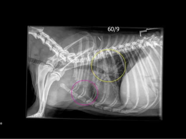
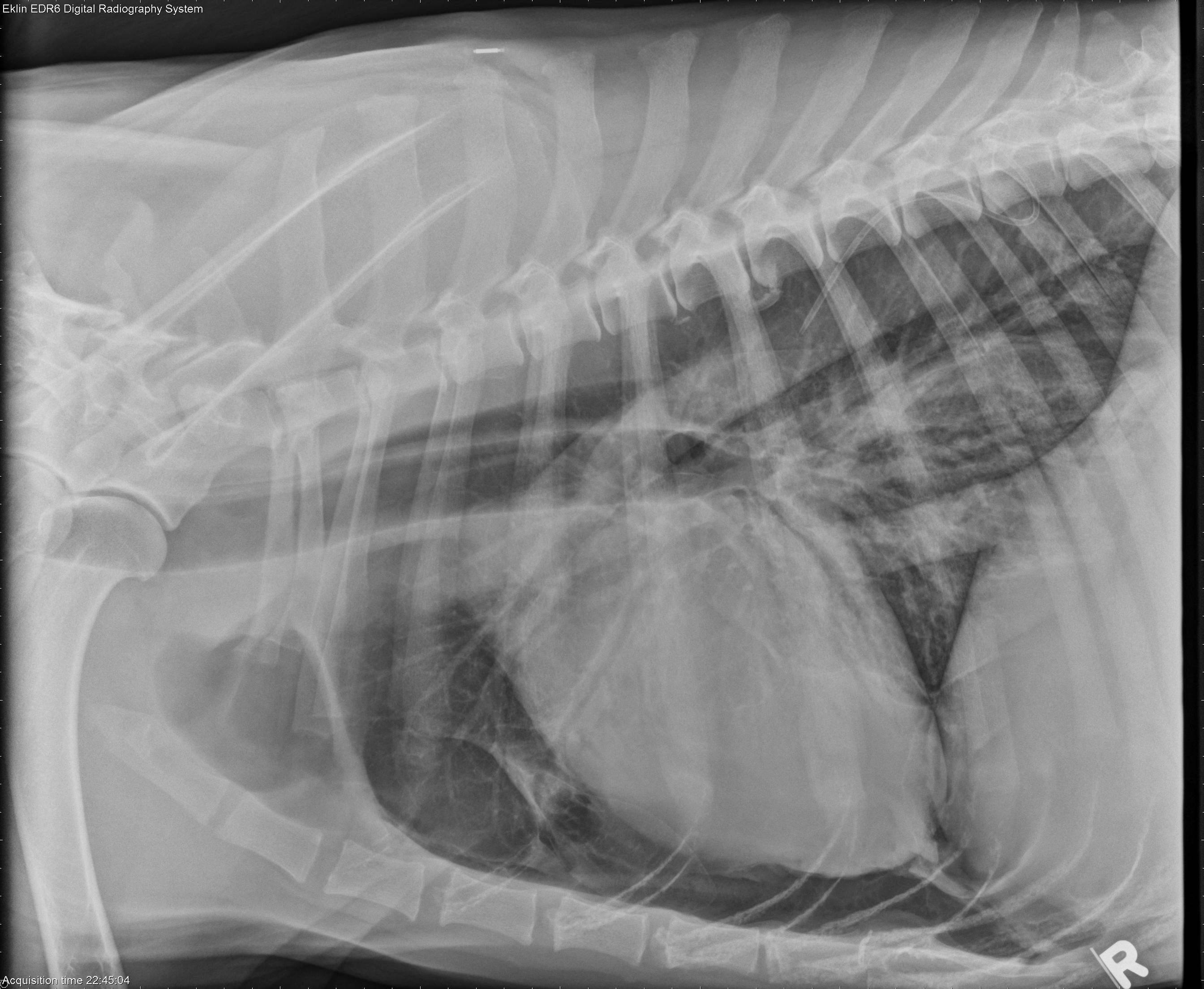
5 Year Old German Shepherd
 www.veterinaryradiology.net
german shepherd
The Radiology Assistant : Mediastinum Lymph Node Map
 radiologyassistant.nl
lymph node mediastinum thoracic mapping classification
Lymph node mediastinum thoracic mapping classification. Esophagectomy with three-field lymph node dissection. 5 year old german shepherd