neck and back anatomy
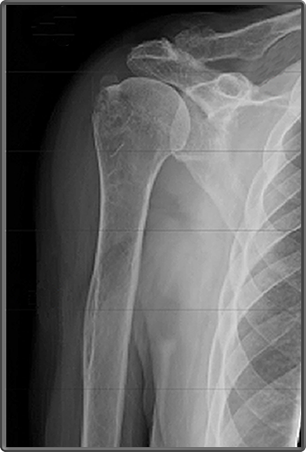
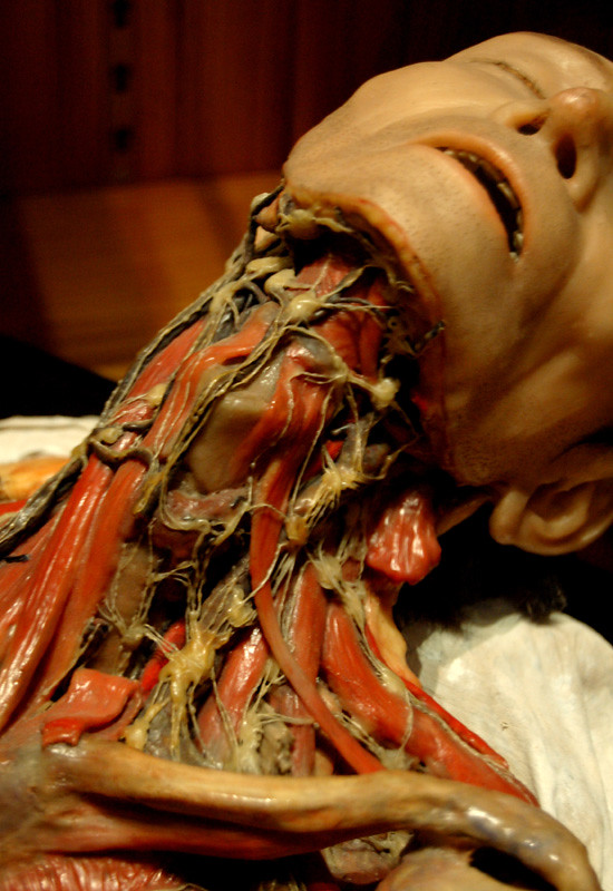
Clinical Anatomy | Radiology | Humerus at Shoulder. 9 Pictures about Clinical Anatomy | Radiology | Humerus at Shoulder : Lymphatic system | 19th-century wax anatomical model showing… | Flickr, Clinical Anatomy | Radiology | Humerus at Shoulder and also Medial plantar muscles of the foot: Anatomy | Kenhub.
Clinical Anatomy | Radiology | Humerus At Shoulder
 www.clinicalanatomy.ca
humerus radiology
What You Need To Know: Anatomy Of The Sciatic Nerve - Spine Surgery San
 spinecenteroftexas.com
sciatic spine
Color X-Ray Of The Cervical Spine Showing Degenerative Disc Disease
 www.pinterest.com
degenerative disease disc cervical spine neck ray ddd pain mri spinal arthritis cord disk showing lumbar nerve symptoms c5 surgery
Lymphatic System | 19th-century Wax Anatomical Model Showing… | Flickr
 www.flickr.com
anatomy wax lymphatic human system lymph nodes anatomical body medical museum neck century 19th scientific sculpture animal showing worlds skin
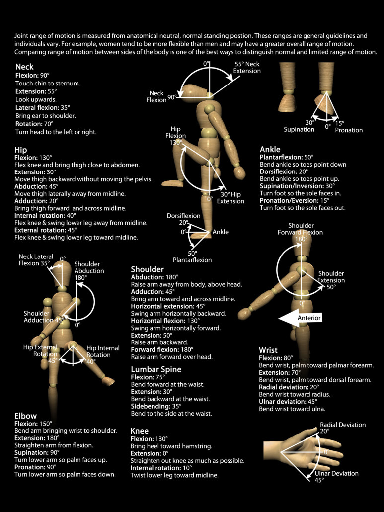
Infographics & Posters - Real Bodywork
 www.realbodywork.com
motion range joint chart degree therapy anatomy rom physical human realbodywork shoulder charts pain ranges massage neck yoga sports reflexology
Suboccipital Muscles - Anatomy | Kenhub
:background_color(FFFFFF):format(jpeg)/images/library/1403/Suboccipital_muscles.png) www.kenhub.com
www.kenhub.com
suboccipital muscles kenhub anatomy
Human Blood Vessels In Neck, Artwork - Stock Image - F009/4015
 www.sciencephoto.com
Parapharyngeal And Retropharyngeal Spaces: Anatomy | Kenhub
:background_color(FFFFFF):format(jpeg)/images/article/en/the-para-and-retropharyngeal-spaces/2tEmtzKqjNappHRcfy7Vg_Pharynx.png) www.kenhub.com
www.kenhub.com
carotid artery common anatomy retropharyngeal parapharyngeal space spaces pharynx dorsal kenhub skull variations anatomical
Medial Plantar Muscles Of The Foot: Anatomy | Kenhub
:background_color(FFFFFF):format(jpeg)/images/article/en/medial-muscles-of-the-sole-of-the-foot/hIK4Yo6xKbeXQRPTZcww_Medial_Plantar_Muscles.png) www.kenhub.com
www.kenhub.com
foot anatomy kenhub muscles plantar medial muscle ankle plantaris sole body diagram human sink 3d nerve tibial study anatomie read
Human blood vessels in neck, artwork. Clinical anatomy. Medial plantar muscles of the foot: anatomy