mouse spinal cord anatomy
Owl-eyes sign (spinal cord) | Radiology Reference Article | Radiopaedia.org. 17 Pics about Owl-eyes sign (spinal cord) | Radiology Reference Article | Radiopaedia.org : | (A) Schematic of the spinal cord, referenced from MRI images of mouse, Image result for mouse spinal cord | Spinal cord, Spinal, Cord and also An Acute Mouse Spinal Cord Slice Preparation for Studying.
Owl-eyes Sign (spinal Cord) | Radiology Reference Article | Radiopaedia.org
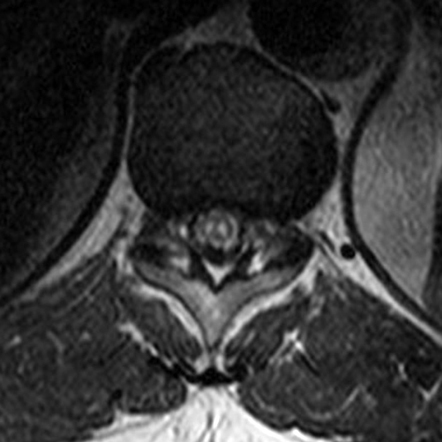 radiopaedia.org
spinal radiology radiopaedia ischemia artery
Filum Terminale | Radiology Reference Article | Radiopaedia.org
 radiopaedia.org
filum terminale spinal enlargement radiopaedia cauda equina radiology
Hematomyelia | Radiology Reference Article | Radiopaedia.org
 radiopaedia.org
spinal cavernoma radiopaedia
An Acute Mouse Spinal Cord Slice Preparation For Studying
 bio-protocol.org
spinal cord mouse slice acute preparation studying
MR Image Gallery – University Of Copenhagen
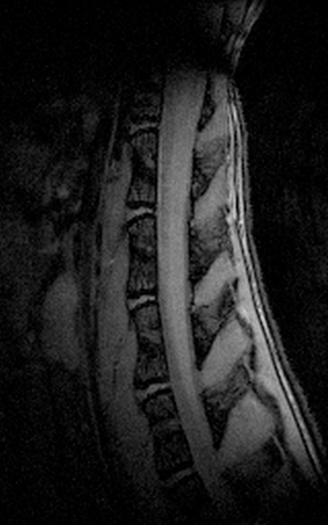 nmr.ku.dk
mr spinal t1wi cord mouse
| (A) Schematic Of The Spinal Cord, Referenced From MRI Images Of Mouse
 www.researchgate.net
dissection referenced c57bl 6j
PPT - Anatomy Of The Kidney & Ureter PowerPoint Presentation - ID:2240729
 www.slideserve.com
lymph kidney ureter anatomy ppt nodes nerve supply drainage renal around artery aortic powerpoint presentation drains lateral
Spinal Cord - Wikipedia
 en.m.wikipedia.org
spinal astrocytes
Mouse. Spinal Cord, Transverse, H+E. (A), (B) Cervical. (C), (D
 www.researchgate.net
transverse dorsal root ventral
Gale Academic OneFile - Document - A Method For Removing The Brain And
histology
Anterior Spinal Artery | Radiology Reference Article | Radiopaedia.org
 radiopaedia.org
spinal radiopaedia artery radiology arterial
Spinal Cord Injury
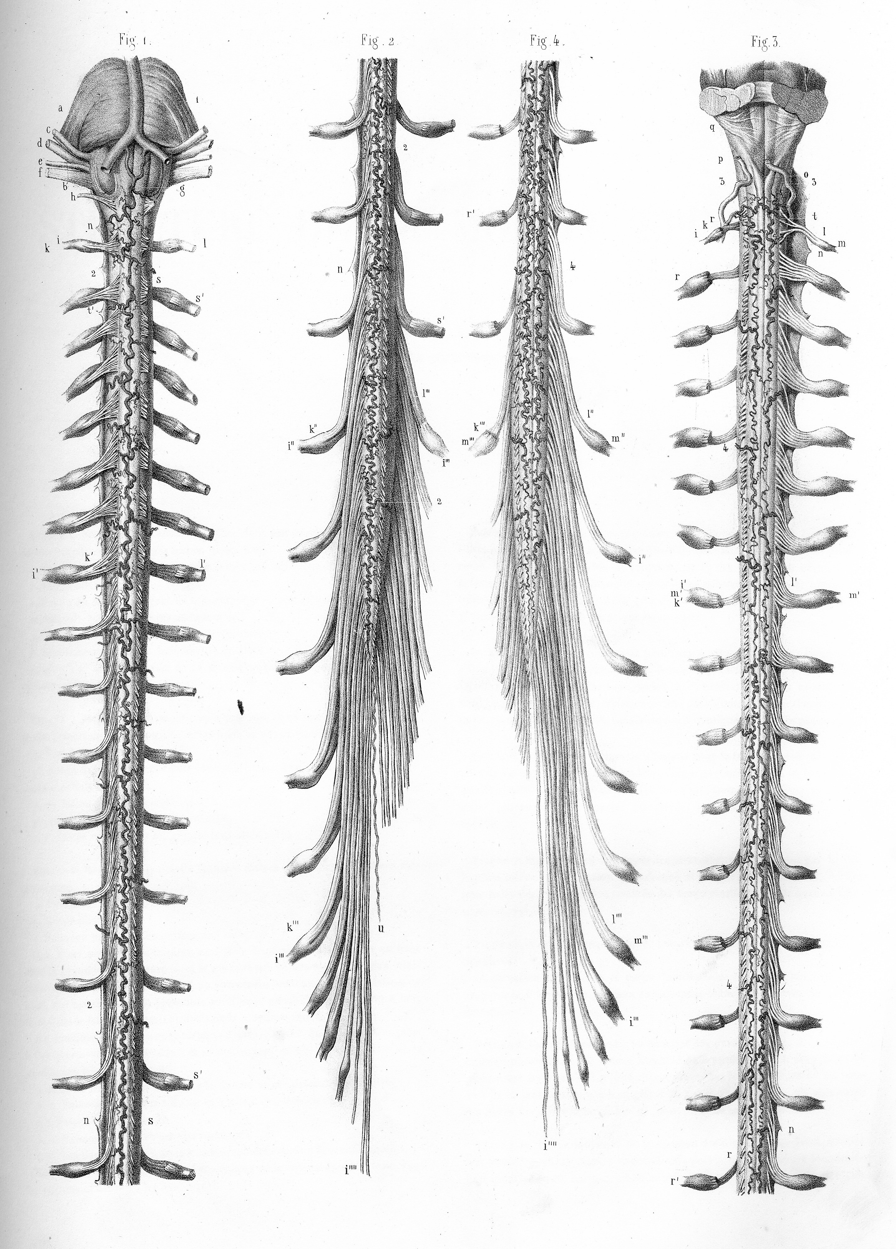 www.animalresearch.info
spinal cord injury drawing info moelle anatomy 2000 épinière schéma tronc sport cérébral du spinale paintingvalley medical
GPI 1510 Sacrum T8 Spine Model
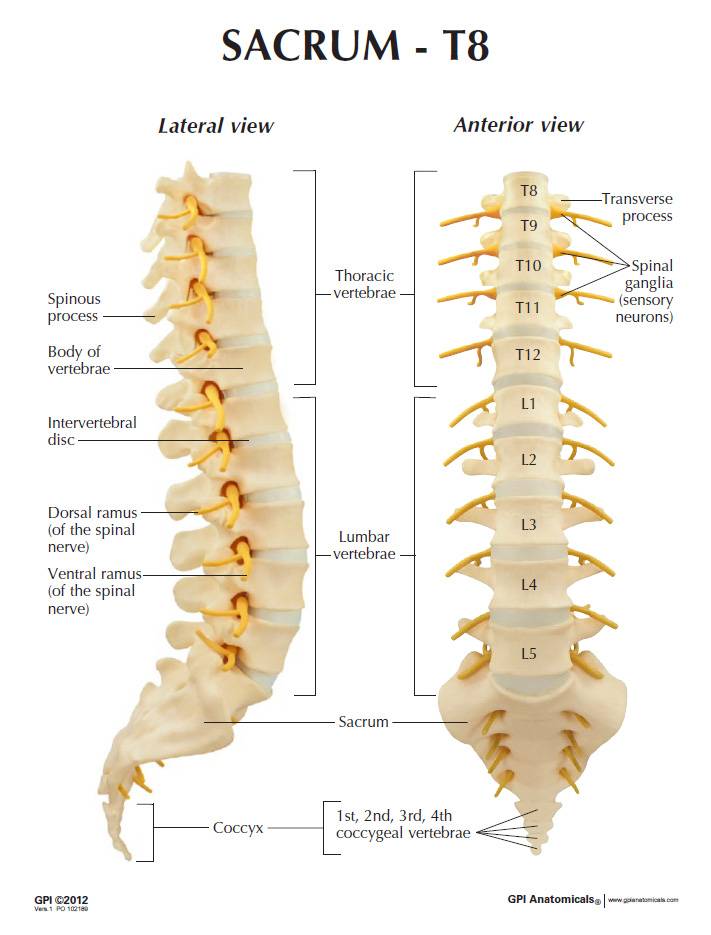 www.universalmedicalinc.com
spine sacrum t8 vertebrae l1 l5 t12 spinal vertebra 1510 anatomical models lfa card
CLARITY For The Mouse Spinal Cord – Spinal Cord Injury Model – SengulLab
 sengullab.com
spinal t9 t8
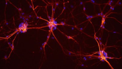
Teaching The Nervous System To Forget Chronic Pain | NOVA | PBS
 www.pbs.org
chronic spinal impulses axons
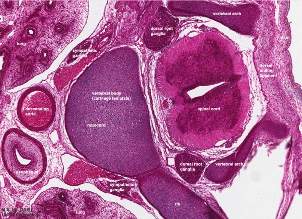
Notochord - Embryology
 embryology.med.unsw.edu.au
spinal cord development embryology vertebra notochord embryo vertebral structure neural skeleton axial human arch embryonic musculoskeletal system unsw med edu
Image Result For Mouse Spinal Cord | Spinal Cord, Spinal, Cord
 www.pinterest.com
spinal cord
Gpi 1510 sacrum t8 spine model. Spinal astrocytes. Spinal radiopaedia artery radiology arterial