liver ultrasound anatomy
Image | Radiopaedia.org. 16 Pictures about Image | Radiopaedia.org : Οι 32 καλύτερες εικόνες του πίνακα LIVER ANATOMY | Ανατομία, LIVER ULTRASOUND ANATOMY 4.wmv - YouTube and also Οι 32 καλύτερες εικόνες του πίνακα LIVER ANATOMY | Ανατομία.
Image | Radiopaedia.org
 radiopaedia.org
cirrhosis radiopaedia liver chronic
106 Best Images About US Liver On Pinterest | Portal, Muscle Tissue And
 www.pinterest.com
liver ultrasound anatomy
The Pancreas | Radiology Key
 radiologykey.com
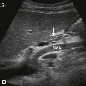
pancreas lobe liver left radiologykey
LIVER ULTRASOUND ANATOMY 4.wmv - YouTube
 www.youtube.com
ultrasound liver anatomy segment rt hepatic 39q own ivc between
Ultrasound Of NORMAL LIVER Part 2 Of 3 - YouTube
 www.youtube.com
liver ultrasound normal
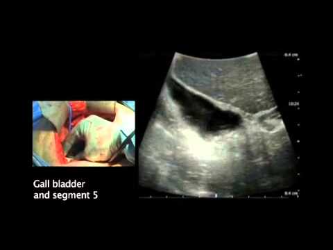
Intraoperative Ultrasound Imaging Of The Liver - YouTube
 www.youtube.com
liver ultrasound intraoperative
Sonography Of The Liver-P2.flv - YouTube
 www.youtube.com
ultrasound liver radiology sonography portal hypertension flv p2 medical
A Gallery Of High-Resolution, Ultrasound, Color Doppler & 3D Images - Liver
 www.ultrasound-images.com
ultrasound lobe beaver elongated ecografia riedels
Exploring The Liver By Ultrasound Along Its Anatomy | Radiology Key
 radiologykey.com
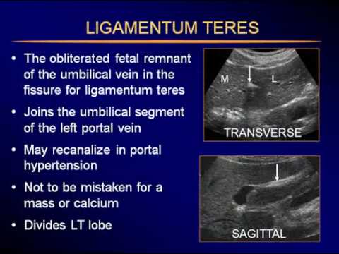
Liver Anatomy And Protocol Basics – Sonographic Tendencies | Ultrasound
 www.pinterest.com
ultrasound
Ultrasound - Radiology Library
 www.vhdissector.com
ultrasound
Liver Atlas: Diagnosis: Cirrhosis
 liveratlas.org
cirrhosis liver findings
Focal Nodular Hyperplasia | Image | Radiopaedia.org
 radiopaedia.org
nodular hyperplasia liver focal sonography gallbladder abnormal radiopaedia steatosis cardiac
Couinaud’s Liver Segments – Sonographic Tendencies In 2021
 www.pinterest.com
liver segments couinaud sonography ultrasound
Οι 32 καλύτερες εικόνες του πίνακα LIVER ANATOMY | Ανατομία
 www.pinterest.fr
Hepatic Angiosarcoma | Image | Radiopaedia.org
 www.radiopaedia.org
angiosarcoma hepatic radiopaedia liver version
Liver anatomy and protocol basics – sonographic tendencies. Liver atlas: diagnosis: cirrhosis. Sonography of the liver-p2.flv