label diagram of human skin
Skin Diagram Not Labeled - img-doppelganger. 13 Images about Skin Diagram Not Labeled - img-doppelganger : Anatomy Skin Diagram Labeled - Diagram Media, Skin Diagram Anatomy And Physiology - Diagram Media and also Skin Diagram Anatomy And Physiology - Diagram Media.
Skin Diagram Not Labeled - Img-doppelganger
 img-doppelganger.blogspot.com
labeled labeling quizlet
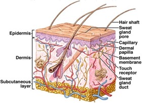
Anatomy Of Human Skin With Labels ストックイラストレーション - Getty Images
 www.gettyimages.co.jp
Endocrine - Other Tissues - Embryology
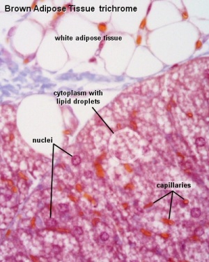 embryology.med.unsw.edu.au
adipose brown tissue tissues histology connective development embryology endocrine fat components cells cartilage edu bat unsw med ossification
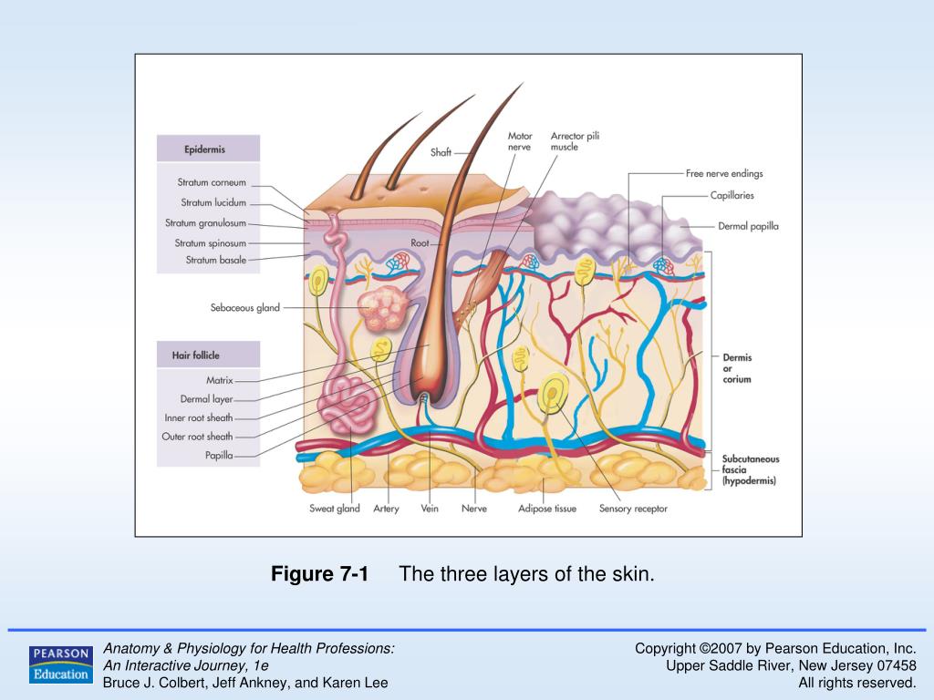
Anatomy Skin Diagram Labeled - Diagram Media
 diagramedia.blogspot.com
labeled labeling
Human Histology For Amateur Microscopists
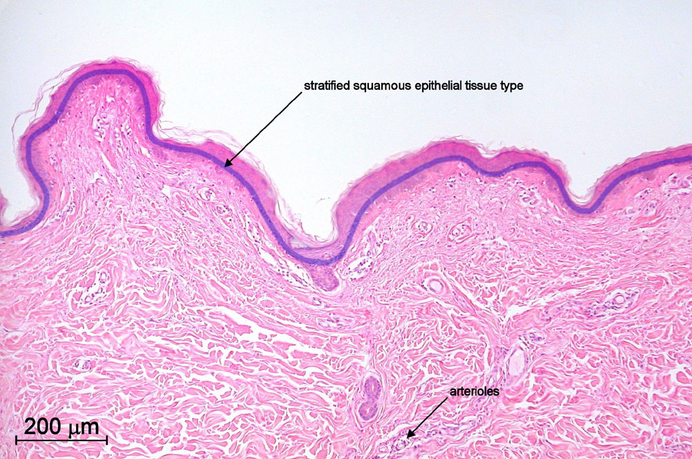 www.microscopy-uk.org.uk
human skin histology 40x thin sample section objective microscopy stained nonpigmented figures larger version mag
Histology Drawings: January 2014
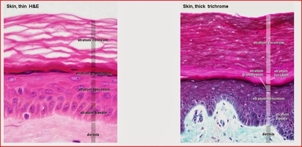 histologydrawings.blogspot.com
skin thin thick histology microscope between light differences epidermis integumentary drawings comparison system specimens
What Is Skin? The Layers Of Human Skin - YouTube
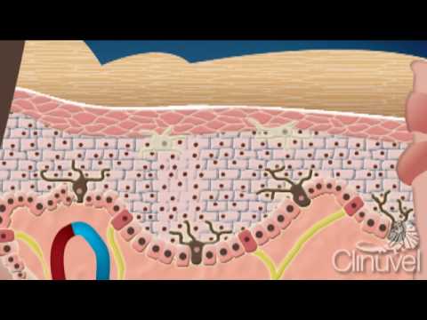 www.youtube.com
human skin layers body organ largest epidermis biology dermis science anatomy sense hypodermis approximately weighing bodyweight cells smell taste touch
Difference Between Sweat And Sebum - MD
 www.majordifferences.com
skin integumentary system integument sweat glands labeled exocrine layers anatomy quizlet human gland layer labels diagram hair sebaceous between label
Free Coloring Pages Of Integumentary System | Integumentary System
 www.pinterest.com
coloring integumentary blank system anatomy human worksheets body diagram skin worksheet labeling structure sheet grade physiology sheets craft science assistant
Skin Diagram Anatomy And Physiology - Diagram Media
 diagramedia.blogspot.com
physiology
Skin Diagram Labeled
 healthiack.com
1122
Cells Under A Microscope By Jaimarie Nelson
 www.haikudeck.com
microscope under cells slide
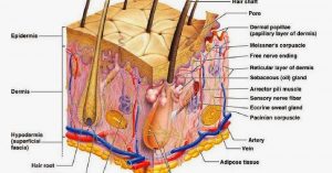
Zaf Naqui | Anatomy
 www.zafnaqui.com
skin subcutaneous anatomy layers administration tissue injection disorders mean does protects against than structure cosmetic ingredients absorbed into deep
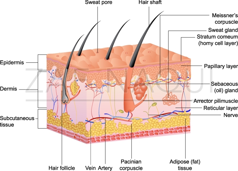
Skin thin thick histology microscope between light differences epidermis integumentary drawings comparison system specimens. Histology drawings: january 2014. Adipose brown tissue tissues histology connective development embryology endocrine fat components cells cartilage edu bat unsw med ossification